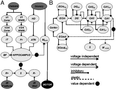
Fig. 2.
Schematic of the regional and functional neuroanatomy of Darwin X. Gray ellipses denote different neural areas. Arrows denote projections from one area to another. (A) Diagram of cortical-hippocampal connectivity. Input to the neural simulation comes from a charge-coupled device camera, wheel odometry, and IR sensors for wall and platform detection. The simulation contains neural areas analogous to visual cortex (V1, V2/4), the inferotemporal cortex (IT), parietal cortex (Pr), head direction units (HD), anterior thalamic nuclei (ATN), motor areas for egocentric heading (MHDG), a value system (S), and positive and negative reward areas (R+, R-). The hippocampus is connected with the three major sensor input streams (IT, Pr, ATN), the motor system (MHDG), and the value system (S). The hippocampus receives rhythmic inhibition from a simulated basal forebrain region (BF). (B) Diagram of connectivity within the hippocampal region. The modeled hippocampus contains areas analogous to entorhinal cortex (ECIN,ECOUT), dentate gyrus (DG), and the CA3 and CA1 subfields. These areas contain interneurons that implement feedback inhibition (e.g., CA3 → CA3FB → CA3) and feed-forward inhibition (e.g., DG → CA3FF → CA3). See supporting information for details.

