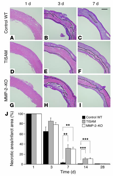
Figure 6.
Histology and morphometry of the LV infarcted myocardium from vehicle-treated control WT, TISAM-treated, and MMP-2–KO mice. Paraffin sections of the hearts obtained from vehicle-treated control WT mice (A–C), TISAM-treated mice (D–F), and MMP-2–KO mice (G–I) on days 1, 3, and 7 were stained with H&E. Necrotic zones are marked by dotted lines. Scale bar: 300 μm. (J) Morphometrical analysis of the LV necrotic area to total infarct area, showing that phagocytic removal of necrotic cardiomyocytes by macrophages was significantly reduced in TISAM-treated and MMP-2–KO mice on days 7 and 14. **P < 0.01; ***P < 0.001.

