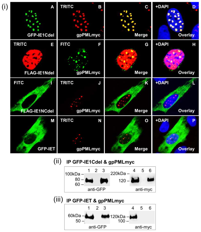
Figure 7. Localization and interaction of gpPML with GPCMV IE1 mutants.
(i) Co-localization of different IE1 truncation mutants and gpPMLmyc in GPL cells analyzed by immunofluorescence. A–D, GFP-IE1Cdel (A) and gpPMLmyc (B) shown separately in the same cell. C merged image for A and B. D overlay of C with DAPI. E–H, FLAG-IE1Ndel (E) and gpPMLmyc (F). G merged image, with H being DAPI overlay. I–L, FLAG-IE1NCdel (I) and gpPMLmyc (J). K merged image, with L being DAPI overlay. M–P, GFP-IET (M) and gpPMLmyc (N). O merged image, with P being DAPI overlay. (ii) GFP-IE1Cdel and gpPMLmyc co-expression and IP. (iii) GFP-IET and gpPMLmyc co-expression and IP. Lanes 1 and 4, total cell lysate of transfected GPL cells. Lanes 3 and 6, IP reactions. Lanes 2 and 5, mock transfected GPL cells. gpPMLmyc, FLAG-IENdel, and FLAG-IE1NCdel detected by primary anti-epitope Ab and secondary anti-mouse IgG FITC or TRITC (immunofluorescence) and anti-mouse IgG-HRP (western blot). GFP-IE1Cdel and GFP-IET detected by fluorescence (cell localization) and specific epitope Ab (western blot).

