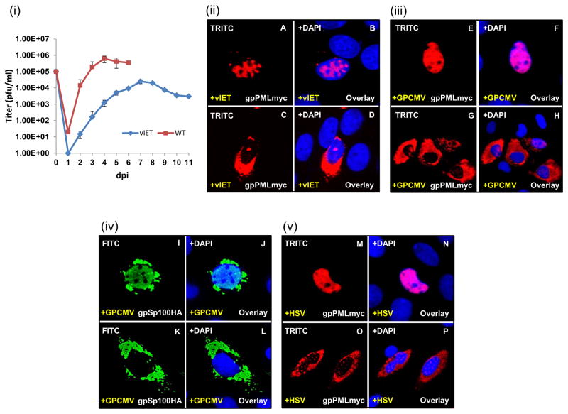
Figure 9. Dispersal of transiently expressed ND10 by WT HSV-1 and GPCMV.
(i) GPL cells were infected with either IET or WT virus (0.1 MOI). Samples taken daily for 6 or 11 days and titrated in duplicate on GPL cells. Results plotted as virus titer versus days post infection (p.i.). (ii-v) GPL cells were infected with HSV-1 or GPCMV (0.1 MOI) prior to transient transfection with the gpPMLmyc expression construct and analyzed for gpPML localization by immunofluorescence. (ii) A–D, expression of gpPMLmyc in the presence of vIET. gpPML redistributes throughout the nucleus (A) and into the cytoplasm (C). Panels B and D are overlays for A and C with DAPI respectively. (iii) E–H, expression of gpPMLmyc in the presence of GPCMV. gpPML redistributes throughout the nucleus (E) and into the cytoplasm (G). Panels F and H are overlays for E and G with DAPI. (iv) I–L, expression of gpSp100HA in the presence of GPCMV causes redistribution of gpSP100 in the nucleus (I) and into the cytoplasm (K). Panels J and L are overlays for I and K with DAPI. (v) M–P, expression of gpPMLmyc in the presence of HSV-1 seen in the nucleus (M) and into the cytoplasm (O). Panels N and P are overlays for M and O with DAPI respectively. gpPMLmyc and gpSp100HA detected by primary anti-epitope Ab and secondary anti-mouse IgG TRITC or anti-rabbit IgG FITC respectively.

