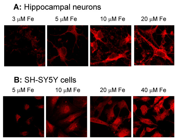
Figure 6.

Immunocytochemistry determination of IREG1. Hippocampal neurons and SH-SY5Y cells, labeled with rabbit anti-IREG1 antibody and with Alexa-546-conjugated goat anti-rabbit IgG, were imaged in a confocal microscope. Shown are representative fields of cells cultured at different iron concentrations. Note the preferentially cytosolic distribution of IREG1.
