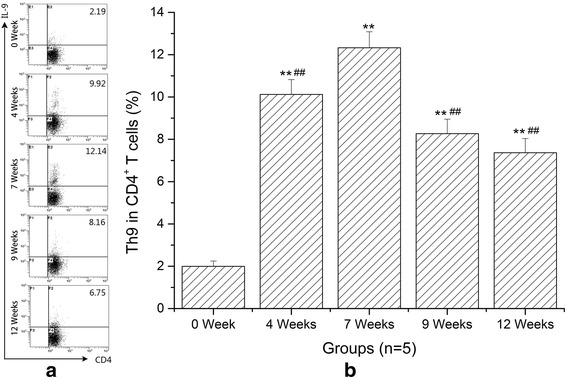
Fig. 4.

Percentages of Th9 cells in total CD4+ T cells from spleens significantly increased in mice with schistosomiasis. a Representative dot plots of Th9 cells by flow cytometry in S. japonicum-infected mice at 0 (NC), 4, 7, 9 and 12 weeks. All of the values were gated on CD4+ cells. The percentages of Th9 cells in CD4+ cells are indicated in the upper right of each chart. b Quantitative changes of Th9 cells in splenic CD4+ T cells at different time points. Data are presented as mean ± SD. **P < 0.01 compared with NC; ## P < 0.01 compared with 7 weeks
