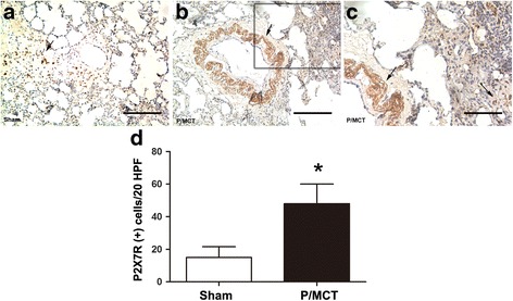
Fig. 1.

IHC staining of P2X7R in the sham group (a) and P/MCT group (b, c) at 4 weeks after MCT injection. Quantification of P2X7R-positive cells per 20 high-power fields (HPFs) (d). Original magnification × 20. Scale bar = 50 μm for all images. IHC: Immunohistochemistry; P/MCT: MCT plus left pneumonectomy
