Fig. 1.

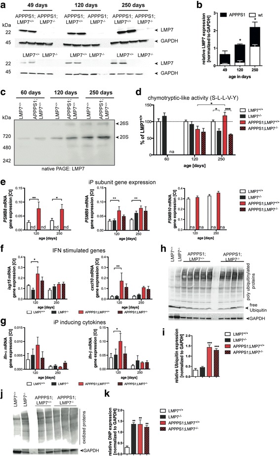
iP subunit expression in the brain is increased upon aging and enhanced in APPPS1 mice. a Western blot analysis of LMP7 expression in brain homogenates of 49d, 120d and 250d old wildtype (LMP7+/+) and APPPS1;LMP7+/+ mice (respective littermate control LMP7−/− and APPPS1;LMP7−/− mice served as a control for antibody specificity) and (b) corresponding densitometric quantification (right panel; n = 3–5 mice per group, (LMP7+/+: 49d vs. 250d; p = 0.0253; LMP7+/+ vs. APPPS1;LMP7+/+: 49d ns; 120d p = 0.0222; 250d p = 0.0065; two-way ANOVA followed by Bonferroni post-tests). c Native PAGE analysis of brain homogenates from LMP7+/+ and APPPS1;LMP7+/+ mice. Proteasome complexes were visualized by western blotting for the iP subunit LMP7. d Total 26S chymotryptic peptide-hydrolyzing activity was analyzed in brain homogenates from LMP7+/+, LMP7−/−, APPPS1;LMP7+/+ and respective littermate APPPS1;LMP7−/− mice using Suc-LLVY-AMC peptide hydrolysis (n = 5 per group; LMP7+/+ vs. LMP7−/−: p = 0.0061; APPPS1;LMP7+/+ vs. APPPS1;LMP7−/−: p = 0.0001; two-way ANOVA followed by Bonferroni post-tests). e-g Quantitative PCR analysis of PSMB8, PSMB9, PSMB10, isg15, cxcl10, Ifn-α and Ifn-β from whole brain tissue of LMP7+/+, LMP7−/−, APPPS1;LMP7+/+ and respective littermate APPPS1;LMP7−/− mice (n = 5 per group; PSMB8 120d: LMP7+/+ vs. APPPS1;LMP7+/+: p = 0.0020; 250d: LMP7+/+ vs. APPPS1;LMP7+/+: p = 0.0162; PSMB9 120d: LMP7+/+ vs. APPPS1;LMP7+/+: p = 0.0041; 250d: LMP7+/+ vs. APPPS1;LMP7+/+: p = 0.0014; isg15 120d: LMP7+/+ vs. APPPS1;LMP7+/+: p = 0.0165; cxcl10 120d: LMP7+/+ vs. APPPS1;LMP7+/+: p = 0.0007; ifn-β: LMP7+/+ vs. APPPS1;LMP7+/+: p = 0.0410; two-way ANOVA followed by Bonferroni post-tests). h Western blot analysis of poly-ubiquitin conjugates in brain homogenates of 120 days old APPPS1;LMP7+/+ and respective littermate APPPS1;LMP7−/− mice as well as age-matched wild-type and LMP7−/− mice and (b) corresponding densitometric quantification (n = 3–5 mice per group; *** p = 0.001, one-way ANOVA followed by Bonferroni post-tests). (j) Western blot analysis of oxidant-damaged proteins in brain homogenates of 120 days old APPPS1;LMP7+/+ and respective littermate APPPS1;LMP7−/− mice, as well as age-matched wild-type and LMP7−/− mice using carbonyl-detection and (k) corresponding densitometric quantification (n = 2–4 mice per group; LMP7+/+ vs. LMP7−/−: p = 0.0096; LMP7+/+ vs. APPPS1;LMP7+/+: p = 0.0066; LMP7+/+ vs. APPPS1;LMP7−/−: p = 0.0023; ** p < 0.01, one-way ANOVA followed by Bonferroni post-tests). (nd = not detected and na = not analyzed)
