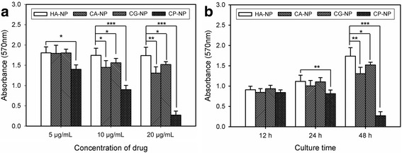
Fig. 6.

In vitro anticancer activity of the drug-loaded CaP nanocomposites on MG-63 cells. The cells were incubated (a) with different concentration of nanocomposites (5–20 μg/mL of drug) for 48 h and (b) with nanocomposites containing 20 μg/mL of drug for different culture time (n = 5). The same amount of SA-NP with CA-NP was used as a reference standard. (p* ˂0.05, p** ˂0.01, p*** ˂0.001)
