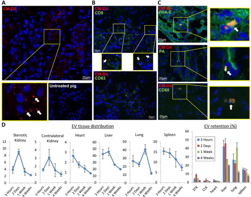
Figure 2. EV membrane fragments were retained within some organelles of renal cells and macrophages.

A: Fragments of red immunofluorescent stained EVs (PKH26, arrows, 40X) were detected in the stenotic kidney 4 weeks after administration (top), whereas untreated stenotic kidneys did not show any fluorescent red signals (bottom). DAPI=blue, nuclei. B: Immunofluorescent co-staining with phaseolus vulgaris erythroagglutinin (PHA-E), peanut agglutinin (PA), and CD68 identified EV fragments within proximal tubular cells, distal tubular cells, and macrophages, respectively. D: Labeled EVs were tracked in frozen sections from the heart, lungs, liver, spleen, and both kidneys, and their retention calculated and expressed as % of injected amount.
