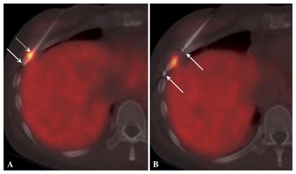
Figure 2.

a) Ga-68 DOTATOC PET/CT guided biopsy. The arrows depict the first biopsy specimen within the needle. The specimen ends were stained green (dotted arrow) and blue (solid arrow) to retain orientation between this image, autoradiography and surgical pathology. b) Ga-68 DOTATOC PET/CT guided cryoablation. A new PET acquisition is used for this fused image. The arrows depict the markers at both ends of active tip of cryoapplicator. The ice ball properly covered the lesion and intended margin during both freeze cycles (not shown).
