
Figure 3.
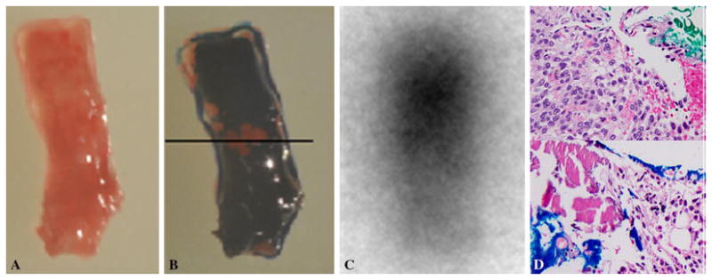
a) color photograph of biopsy specimen in an orientation close to figure 2a. b) The end of the specimen close to the tip of needle (solid arrow on figure 2a) is stained blue at the bottom of this image and the other end (dotted arrow on figure 2a) is stained green on top of the image. The black line depicts the two stained ends. c) autoradiography of biopsy specimen in the same orientation shows intense activity toward the end stained green. d) Select images of each end of specimen show tumor cells on the end stained green and normal marrow elements on the blue stained end. The green end contains monomorphic spindle cells with no atypia or abnormal mitotic activity representing phosphaturic mesenchymal tumor mixed connective tissue variant. The findings on Ga-68 DOTATOC PET/CT guided biopsy, autoradiography and surgical pathology are concordant.