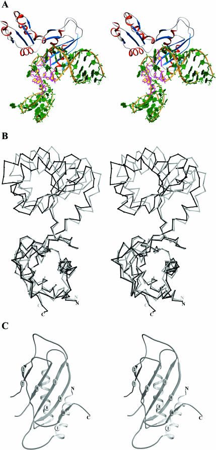
Figure 2.
(A) Stereo view of the L1–mRNA complex. The ribose-phosphate backbone is in gold, bases are in green, β-strands in blue and α-helices in red. Conserved nucleotides are shown in magenta and yellow. (B) Superposition of the structures of the isolated MjaL1 protein (gray) and MjaL1 from the present complex (black) with least squares minimization of differences in Cα atom coordinates of domain I. (C) Stereo view of the MjaL1 domain I.

