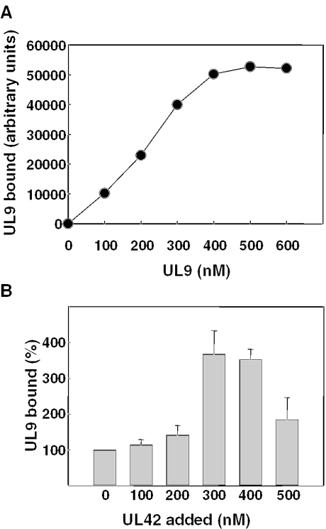
Figure 4.
Equilibrium binding of UL9 to ss DNA. (A) Increasing concentrations of UL9 were incubated with 200 nM 38mer biotinylated at the 5′ end, bound protein was cross-linked to DNA with 1% formaldehyde, and the DNA complexes were isolated using streptavidin beads as described in Materials and Methods. The amount of UL9 in the complexes was determined by phosphorimage analysis of immunoblots using UL9-specific antibody and [125I]protein A and plotted as a function of initial UL9 concentration. (B) A limiting concentration of UL9 (200 nM) was incubated with 200 nM biotinylated 38mer followed by the addition of increasing concentrations of UL42 as indicated. DNA–protein complexes formed at equilibrium were cross-linked, isolated, and the UL9 bound was quantified as indicated above. Within each experiment, the amount of UL9 bound to DNA in the presence of UL42 was normalized to that bound in the absence of UL42 (set at 100%). Results shown represent the mean normalized values obtained (± standard error) for three independent experiments.

