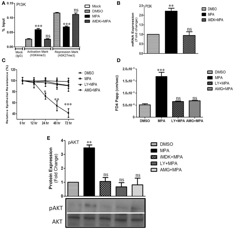
Figure 1.
Influence of MPA on PI3K/AKT pathway and PI3K/AKT dependent modulation of TJs permeability. (A) ChIP-qPCR of gene activation mark (H3K4me3) and repression mark (H3K27me3) at the promoter region of PI3Kgamma gene in Caco-2 cells monolayer treated with either DMSO (Control) or MPA alone or in combination with Midkine inhibitor. (B) The mRNA expression of PI3K gene in Caco-2 cell monolayers treated with either DMSO (Control) or MPA alone or with Midkine inhibitor. GAPDH was used as a house keeping gene. (C) The influence of PI3K pathway inhibition on TJs permeability of Caco-2 monolayer treated with MPA. TEER was measured at 0, 12, 24, 48, and 72 h time points after DMSO (Control) or MPA with or without PI3K inhibitors (LY or AMG). (D) Paracellular FD4 dye flux assay results of Caco-2 monolayers treated with either MPA alone or in combination with PI3K inhibitors (LY or AMG). (E) Representative Western blot and densitrometric analysis of AKT/pAKT in Caco-2 cell monolayers treated with either MPA alone or in combination with Midkine inhibitor (iMDK), or PI3K inhibitors (LY or AMG). Data are shown as mean band density of pAKT relative to total AKT expression. (A–D) Statistically significant differences analyzed by ANOVA with Bonferroni post-test for multiple comparisons are indicated by *P < 0.05, **P < 0.01, ***P < 0.001. ns, non-significant. Error bars represent ± SEM (n = 3).

