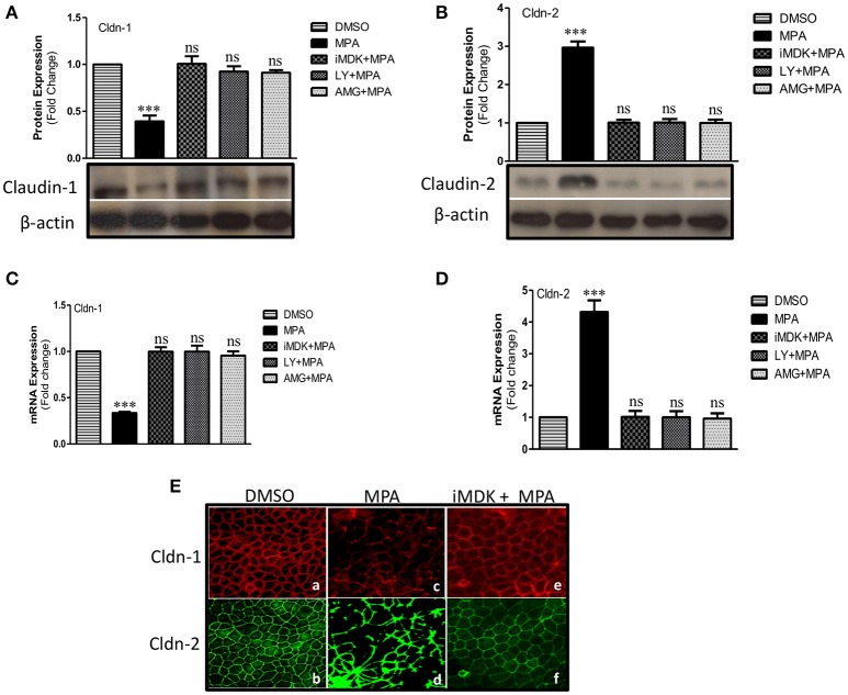
Figure 3.
Influence of Midkine mediated PI3K pathway on TJ structural proteins (Cldn-1, -2) in MPA treated Caco-2 cells. (A,B) Western blot analysis of TJ proteins (Cldn-1, -2) in Caco-2 cell monolayers treated with MPA alone or in combination with Midkine/PI3K inhibitors (iMDK/LY/AMG). Immunoblots were probed with antibodies against TJ proteins (A) Cldn-1 and (B) Cldn-2. β-actin was used as a loading control. (C,D) qRT-PCR analysis of Cldn-1 and -2 gene expression in Caco-2 cell monolayers treated with MPA or Midkine/PI3K inhibitors (iMDK/LY/AMG). GAPDH was used as a house keeping gene and differences between groups were analyzed by ANOVA with Bonferroni post-test. The values were expressed as means ± SEM (n = 3). ***P < 0.001, or ns, non-significant when compared with control cells. (E) Differentiated/polarized monolayer of Caco-2 cells were treated with either DMSO, MPA alone or in combination with Midkine inhibitor (iMDK) for 72 h, fixed, permeated, and stained for Cldn-1 and Cldn-2. The distribution of Cldn-1 and Cldn-2 in differentiated and polarized Caco-2 monolayer exposed to DMSO (a,b), or MPA (c,d) or iMDK+MPA (e,f) is shown.

