Figure 1.
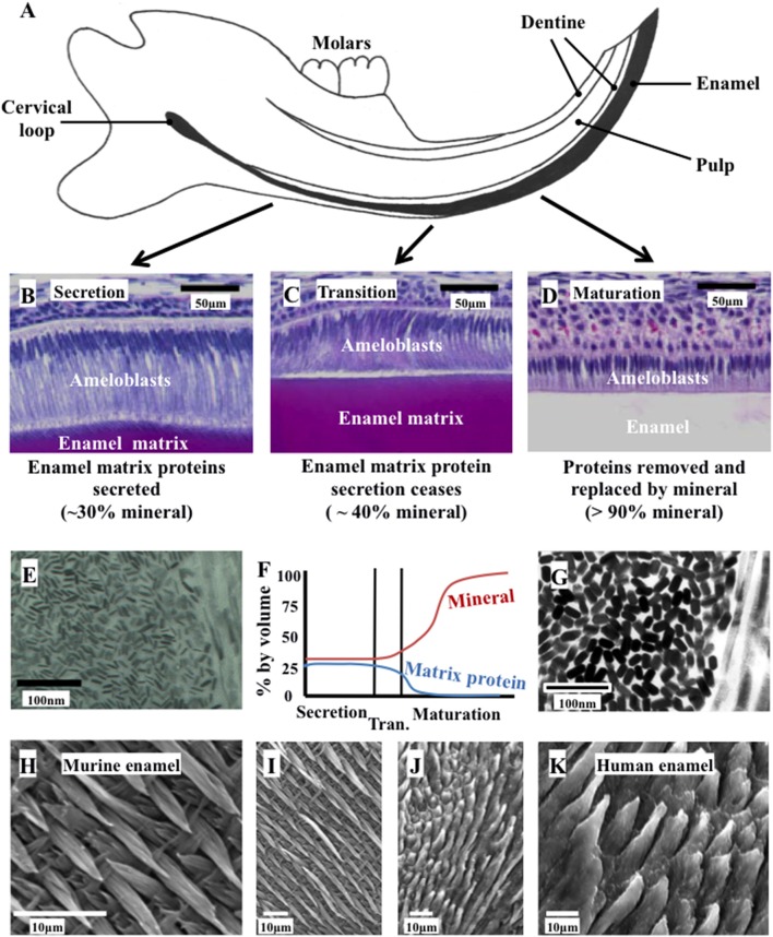
Ameloblast morphology, crystal development, and the final structure of the enamel. (A) Schematic cross section of the murine incisor. (B–D) Histology of the murine incisor. (B) During secretion, ameloblasts exhibit an elongated morphology with a cellular extension (the Tomes' process); (C) during transition the Tomes' process degenerates and the ameloblasts begin to reduce in height; (D) during maturation, the ameloblasts remove nearly all protein from the developing enamel and supply mineral ions to support crystallite growth; (E) immature enamel crystalites form during secretion by growth in their long axis; (F) by the end of secretion, the developing enamel is around 30% mineral and 25% matrix protein, with the remainder tissue fluid. By the end of maturation, the enamel is nearly 100% mineral; (G) the enamel crystallites grow in width and thickness during enamel maturation; (H) and (I) murine enamel has a decussating arrangement of enamel prisms; (J) and (K) human enamel is also arranged in a prismatic structure. Elements from this figure have been adapted from previously published figures and we acknowledge the following publications and publishers for the elements specified: Panels (A–D) were previously published by Barron et al. (2010). Panel (E) was previously published by Robinson (2014).

