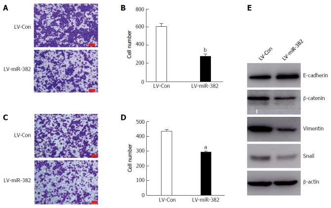
Figure 5.

Overexpression miR-382 inhibited migration, invasion and epithelial-mesenchymal transition in Eca109 cells. A: Representative photomicrographs showed migrated cells stained with crystal violet after post-culture for 24 h in the transwell migration assay. The bars represent 200 μm; B: The migrated cells in five different fields were counted and the Y-axis represents the migrated cell number from three independent experiments. Data is presented as mean ± SD, bP < 0.01; C: Representative photomicrographs showing invaded cells stained with crystal violet after post-culture for 24 h in the transwell invasion assay. The bars are 200 μm; D: The invaded cells in five different fields were counted and the Y-axis represents the invaded cell number from three independent experiments. Data are presented as mean ± SD, aP < 0.05; E: Western blot analysis of the expression of epithelial marker E-cadherin, and mesenchymal markers β-catenin, vimentin and snail in Eca109 cells infected with LV-miR-382 or LV-Con(c). β-actin served as a loading control.
