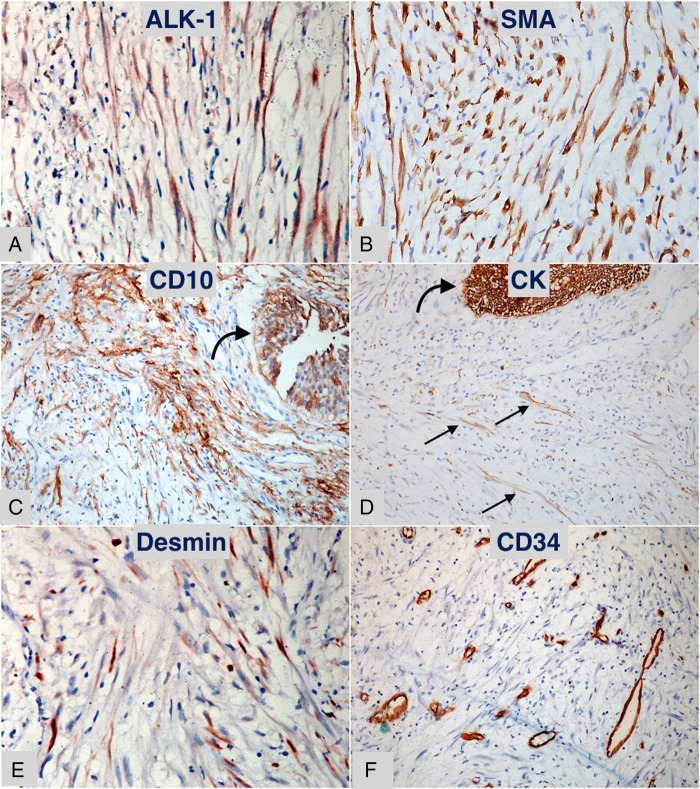
Figure 3.
Diaminobenzidine chromogen stained immunohistochemical sections of the lesion. (A) Anaplastic lymphoma kinase-1 positive myofibroblastic cells (×400). (B) Diffuse positivity for smooth muscle actin in myofibroblastic cells (×400). (C) CD10 positivity in urothelium (curved arrow) and underlying fascicles of myofibroblasts (×200). (D) Cytokeratin positive urothelium (curved arrow) with scattered positive myofibroblasts (small straight arrows) (×200). (E) Scattered focal desmin positive myofibroblasts (×400). (F) CD34 stain highlighting vascular channels, neighbouring tumour cells are negative (×200).

