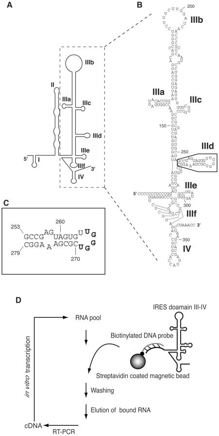
Figure 1.
(A) Schematic structure of the HCV IRES RNA [adapted from (35)]. Structural domains are shown as I–IV. (B) Detailed nucleotide sequence of domain III and IV RNA used in this study. Boxed region indicates domain IIId. (C) Secondary structure of domain IIId. In this report, the sequence bound to the consensus sequence of aptamer is shown in bold letters. (D) Schematic of the in vitro selection procedure to isolate RNAs that bind to the HCV IRES.

