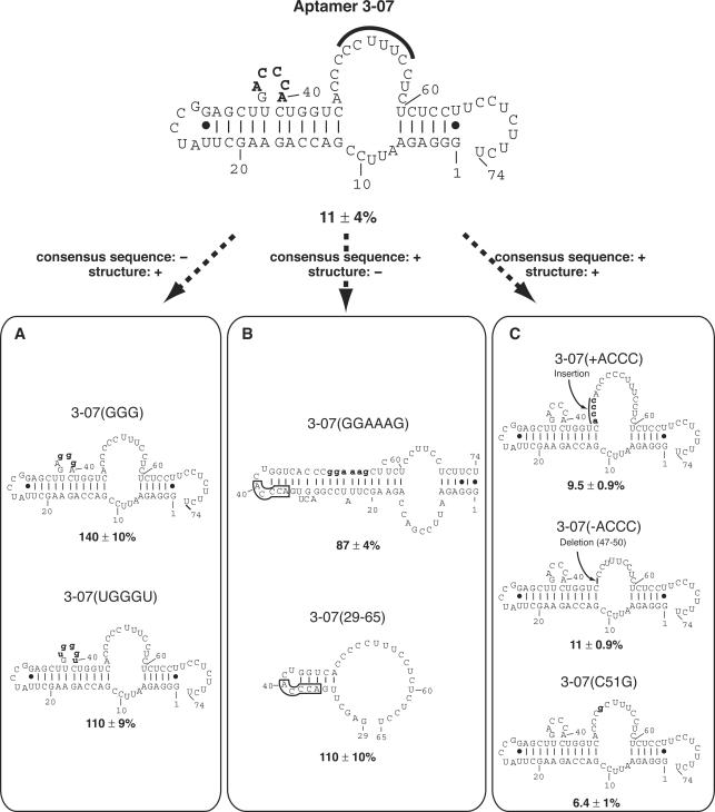
Figure 5.
Structure–inhibition relationships of aptamer 3-07 and its mutants in vitro. Relative luciferase activity in the absence of aptamer was set at 100% and is shown below each structure. Numbering is based on the original aptamer 3-07. (Top) Secondary structure of aptamer 3-07. The consensus sequence is shown in bold letters. The sequence CCUUUUC that is complementary to the stem region of IRES domain IIId is marked by bold line. (Bottom) Mutants are classified by sequence and structure; the consensus sequence and secondary structure described as retaining (+) or not (−). All secondary structures were drawn by the Mulfold program and numbering is based on the original aptamer 3-07. In (B), the boxed regions indicate the consensus sequence. Mutated sequences are indicated by bold and lowercase letters.

