Fig. 3.

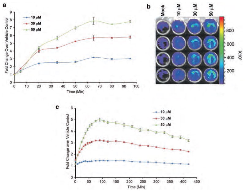
Firefly complementation-based live cell assay for noninvasive monitoring of ATM kinase activity. (a) D54 cells stably expressing the ATM kinase activity reporter (ATMR) were plated in black-walled 96-well plates (5000 cells/well). Cells were incubated with mock (DMSO) or an increasing concentration of ATM kinase inhibitor KU-55933, which increased the signal in a dose-dependent way. (b) A representative image of the bioluminescence acquired in response to various concentrations of KU-55933 is shown. ROI in a grid is created and overlaid in the pseudocolored image to quantitate the photons emitted. Scale bar shows photon flux in pseudocolor with blue as the lowest and red as the highest counts. (c) D54-ATMR cells plated in white-walled 96-well plates and treated as described above and read on Envision plate reader
