Figure 2.

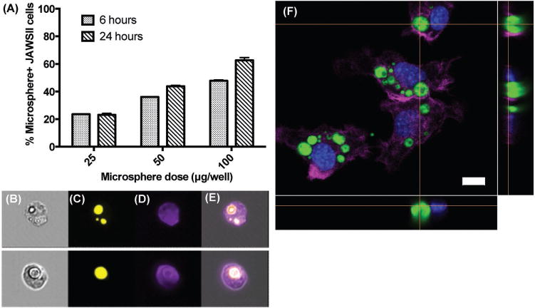
Internalization of self-healing PLGA microspheres by dendritic cells. A) Flow cytometry analysis of JAWSII dendritic cells treated with different doses of rhodamine 6G-labeled microspheres for 6 or 24 h. Columns show the percent of gated events containing cells with associated microspheres. Data represent mean ± SEM (n = 3). Representative ImageStreamX images showing B) bright-field image of JAWSII cells with associated microspheres; C, D) fluorescence images of the microspheres and JAWSII cells, respectively; and E) overlay of images. F) Confocal microscopy image showing JAWSII dendritic cells that internalized rhodamine-labeled self-encapsulating microspheres (green) after 24 h of incubation. Actin filaments were stained with Alexa Fluor 647-phalloidin (violet) and nuclei were stained with DAPI (blue). Scale bar represents 10 μm.
