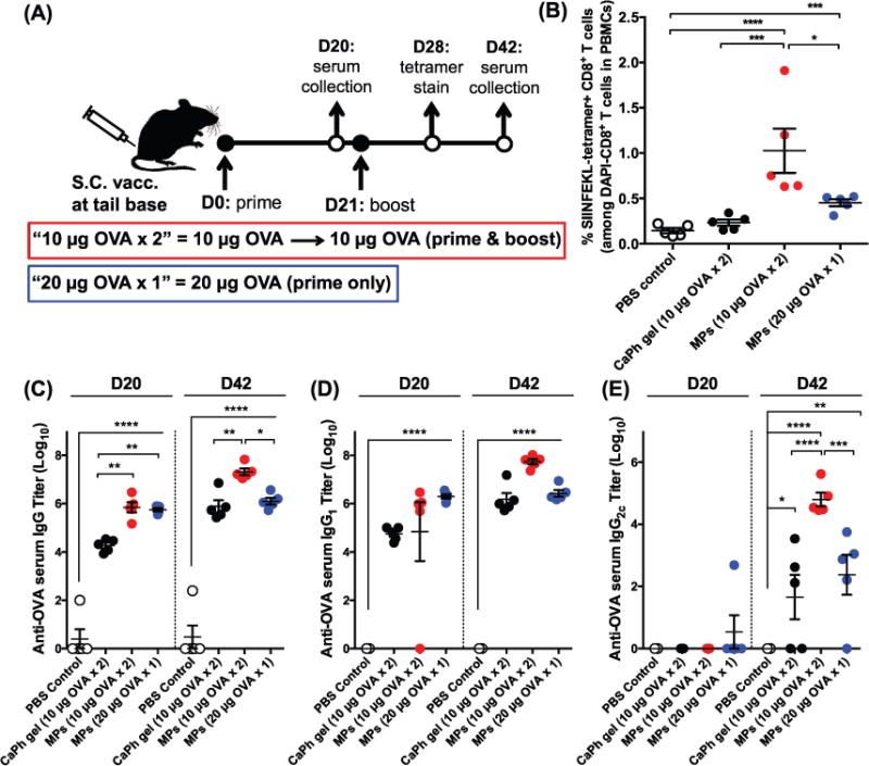
Figure 3.

Cellular and humoral immune responses elicited by self-healing PLGA microspheres in vivo. A) Shown are the vaccine doses and regimen. Naïve C57BL/6 mice were administered subcutaneously at tail base on days 0 and 21 with 10 μg of OVA either admixed with CaHPO4 adjuvant gel or formulated with CaHPO4 adjuvant gel in self-healing PLGA microspheres (10 μg × 2). A group of mice was immunized on day 0 with a prime-only administration of 20 μg of OVA formulated with CaHPO4 adjuvant gel in self-healing PLGA microspheres (20 μg × 1). B) Shown are the percentages of SIINFEKL-tetramer + CD8α + T cells among total CD8α + T cells in PBMCs on day 28. C–E) Serum anti-OVA antibody titers were measured on day 20 (prime response) and day 42 (boost response). Shown are OVA-specific serum C) IgG, D) IgG1, and E) IgG2C titers. Data were fit using a 4-parameter curve, and titers were calculated by solving for the inverse dilution factor resulting in an absorbance value of 0.5. Data represent mean ± SEM (n = 5). All groups were compared using one-way ANOVA (B) or two-way ANOVA (C–E), followed by Bonferroni’s post-test (*P < 0.05, **P < 0.01, ***P < 0.001, and ****P < 0.0001).
