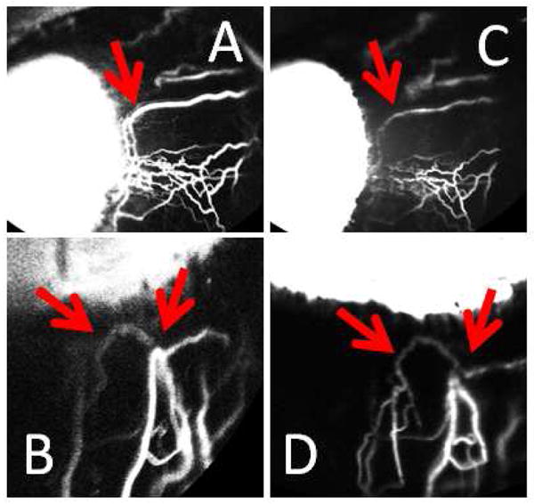
Figure 6. Sequential Aqueous Angiography with ICG and Fluorescein Demonstrated Similar Patterns.

In one eye (NHP-C), sequential aqueous angiography was performed with ICG (A/B) followed by fluorescein (C/D). Like results in post-mortem human eyes, similar patterns were observed between the two dyes (red arrows).
