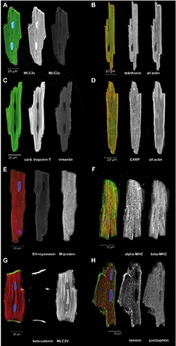
Figure 3.

Freshly isolated ARVM were immediately fixed and immunostained for the indicated proteins. A-H) Overlay images of single optical sections are shown in color, some including DAPI staining for DNA in blue, followed by separate images in greyscale with the green channel in the middle and red channel on the right.
