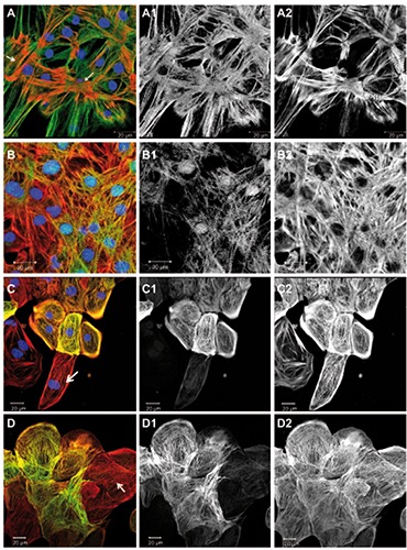
Figure 5.

Developmentally regulated sarcomeric proteins. Overlay images including DAPI are shown on the left, followed by corresponding green and red channel images in greyscale. Images A and B were taken at higher magnification to resolve individual sarcomers, while C and D show larger groups of cells, where labelled proteins appeared in a sub-population of cells. A) Cells were immunostained for telethonin (green), all actin (red), and DNA (blue); arrows point at foci of sarcomeric organization. B) Cells were immunostained for CARP (green), all actin (red), and DNA (blue). C) Cells were immunostained for cardiac troponin-I (green), all actin (red), and DNA (blue); the arrow points at a cardiomyocyte negative for troponin-I. All actin labeled all the cells (C2). D) Cells were immunostained for alpha-smooth muscle actin (green) and all actin (red); the arrow points at a cardiomyocyte negative for alpha-smooth muscle actin and showing sarcomeric striation in the actin channel; actin was observed in all cells (D2).
