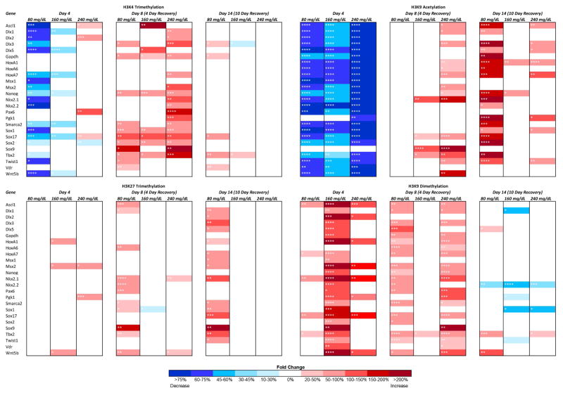
Fig. 1.
Dose-dependent and histone modification-specific changes in chromatin structure. Murine embryonic stem cells were cultured in varying concentrations of ethanol (80 mg/dL, 160 mg/dL, or 240 mg/dL) for four days, followed by a no-ethanol recovery period for ten days. Samples were taken at Day-4, Day-8, and Day-14 and analyze for enrichment of H3K4me3, H3K9ac, H3K27me3, and H3K9me2 using chromatin immunoprecipitation. The enrichment of the indicated post-translational histone modifications was then analyzed within the promoter regions of candidate genes using qPCR. The heat map represents fold change compared to the control group. Within the three separate biological replicates (N = 3), three ChIPs were performed, and two qPCR replicates performed on each independent ChIP. * p<0.05, ** p<0.01, *** p<0.001, **** p<0.0001.

