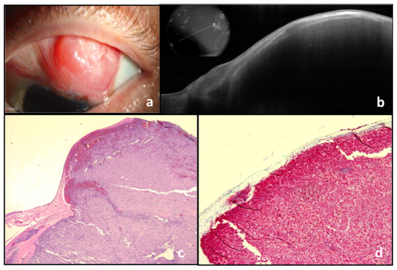
Figure 3. A 56 year old male with amelanotic melanoma of the left eye.

a. Slit lamp photograph of a conjunctival lesion, extending to the limbus. The mass is predominately amelanotic with the exception of small area of pigment at the limbus.
b. AS-OCT showing a thin epithelium overlying a large and elevated subepithelial mass with shadowing of the underlying tissue. The OCT is consistent with the appearance of a conjunctival melanoma.
c. Atypical basophilic cells with prominent nucleoli present within the substantia propria (Original magnification x40)
d. Melan-A with red chromogen highlights the tumor cells within the substantia propria (Original magnification x100), consistent with conjunctival melanoma
