Figure 8.
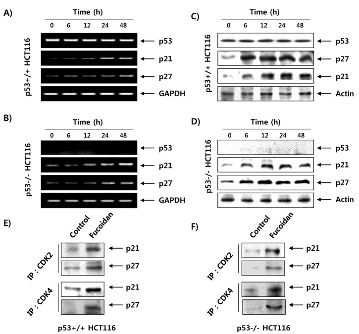
Induction of CDKIs and enhanced association of CDKIs with CDKs by fucoidan in HCT116 cells. (A,B) After treatment with 150 μg/mL of fucoidan for the indicated times, total RNA was isolated and reverse transcribed. The resulting cDNAs were then subjected to PCR with the indicated primers, and the reaction products were separated on 1.5% agarose gel and visualized by EtBr staining. (C,D) The cell lysates were prepared, and equal amounts of total cell lysates were subjected to SDS–PAGE, transferred, and probed with the indicated antibodies. GAPDH and actin were used as internal controls for the reverse transcription (RT)–PCR and Western blot assays, respectively. (E,F) The cells were treated with 150 μg/mL of fucoidan for 24 h, and then total cell lysates (500 μg) were prepared and immunoprecipitated with the anti-CDK2 or anti-CDK4 antibodies. The immunocomplexes were separated on 12% SDS–polyacrylamide gels, transferred onto membranes, probed with anti-p21 or anti-p27 antibodies, and visualized using an ECL detection system.

