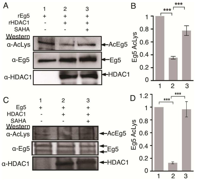
Figure 4.
Eg5 is deacetylated by HDAC1. A) Chemically acetylated full length recombinant Eg5 was incubated in the absence or presence of recombinant HDAC1 and SAHA for 2 hr at 37°C and analyzed by SDS-PAGE and western blotting with acetyl lysine (top) and a combination of Eg5 and HDAC1 (bottom) antibodies. B) Quantification of the Ac-Lys western blot from part A. Mean and standard error of three independent trials are shown (Figures S8A–D). Quantification of the Eg5 blot as a load control is shown in Figures S8E–F. ***p<0.0001. C) Eg5 was immunoprecipitated from T-Ag Jurkat cells (lane 1) and incubated with immunoprecipitated HDAC1 (lane 2) in the presence or absence of 3 mM SAHA (lane 3) for 1 hr at 30°C, followed by SDS-PAGE separation and immunoblotting with acetyl lysine (top), Eg5 (middle), and HDAC1 (bottom) antibodies. D) Quantification of the Ac-Lys western blot from part C. Mean and standard error of three independent trials are shown (Figures S9A–D). Quantification of the Eg5 blot as a load control is shown in Figures S9E–F. ***p<0.0001

