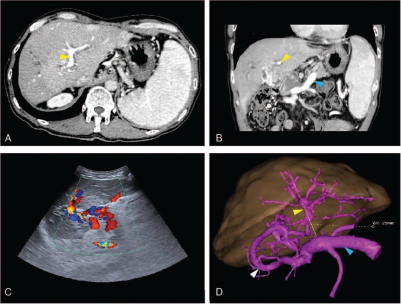
Figure 1.

Preoperative enhanced CT scan (A, B) and Doppler US images (C) showed a patent LPV (yellow arrow), SV (blue arrow) and the confluence of the SMV and SV (D). On 3D reconstruction, an obvious varix presented (white arrow). The distance between the LPV and SV, measured to facilitate surgery, was 69.25 mm (yellow line) (D). CT = computed tomography, LPV = left portal vein, SMV = superior mesenteric vein, SV = splenic vein, 3D = three dimensional, US = ultrasound.
