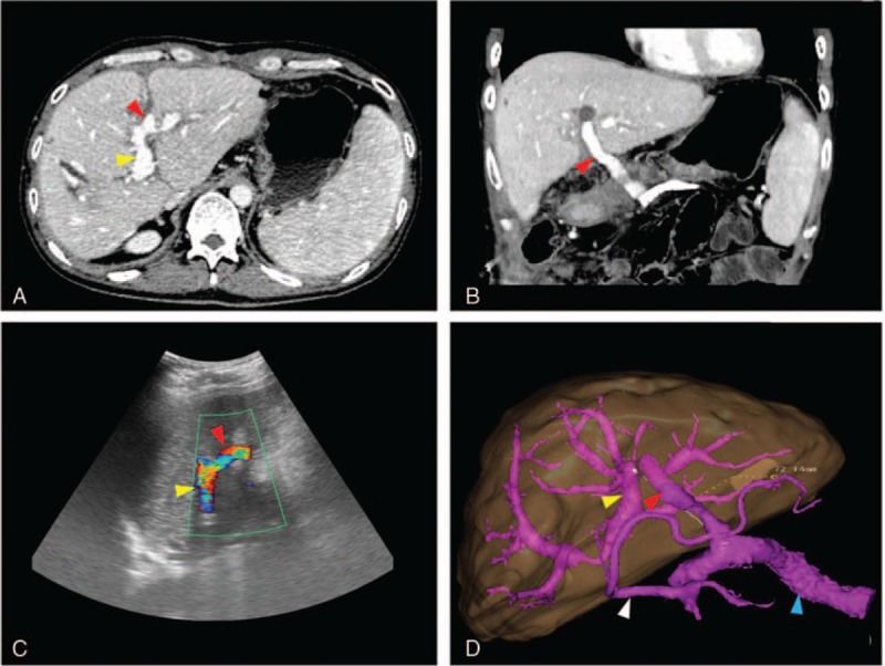
Figure 3.

Postoperative enhanced CT scan (A, B) and Doppler US images (C) showed a patent LPV (yellow arrow), MRB (red arrow) and SV (blue arrow); the LPV became dilated and the varix improved (white arrow). On 3D reconstruction, the length of the bypass was shown to be 72.14 mm (D). CT = computed tomography, LPV = left portal vein, MRB = Meso-Rex bypass, SMV = superior mesenteric vein, SV = splenic vein, 3D = three dimensional, US = ultrasound.
