Figure 9.

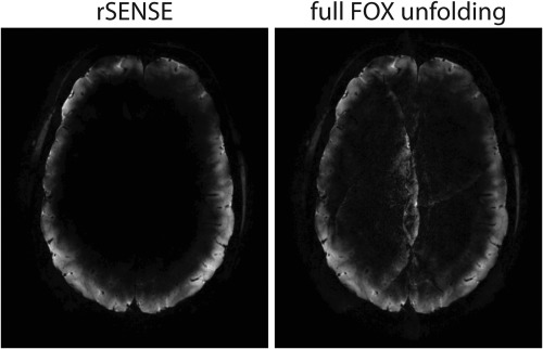
PI accelerated (AF = 3 × 3), T2* weighted, transverse reconstructed images of rFOX shape 3, exciting the outer cortex. The images were reconstructed using rSENSE (left) and using the coil sensitivities of the full FOX (right).

PI accelerated (AF = 3 × 3), T2* weighted, transverse reconstructed images of rFOX shape 3, exciting the outer cortex. The images were reconstructed using rSENSE (left) and using the coil sensitivities of the full FOX (right).