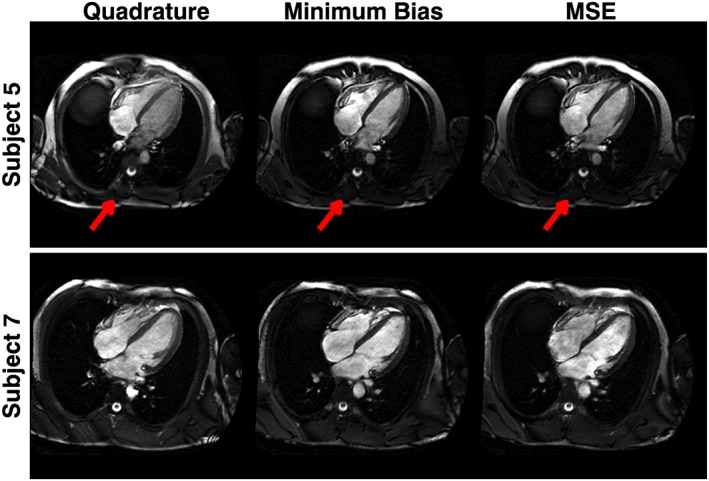
Figure 8.

Full field‐of‐view images for two subjects. The radiofrequency (RF) shimmed solutions (both minimum bias and minimum squared error, MSE) result in low B 1 + in the posterior part of the torso, resulting in low signal (arrows). The image quality for the heart is uncompromised
