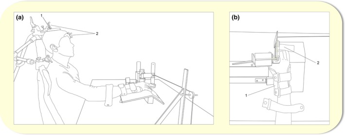
Figure 2.

(a) Illustration of the experimental set‐up during the subTMS and paired‐pulse TMS protocol in a sagittal plane. The stimulator coil (1) was mounted with a coil tracker (2), and markers were attached to the participant's forehead (2) as shown in the picture. (b) Illustration of a closer look of the experimental set‐up used during all experimental sessions in a transverse plane. The arm was held in a pronated position by a splint (1) so that the finger movements were restricted to only allow abduction and adduction of the right index finger. Electromyographical (EMG) electrodes were placed on the right first dorsal interosseous (FDI) (not illustrated). TMS, transcranial magnetic stimulation.
