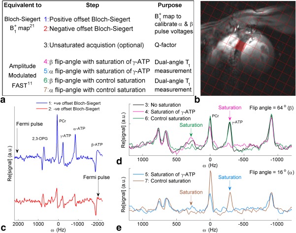
Figure 1.

(a) Summary of Bloch‐Siegert four‐angle saturation transfer protocol steps. (b) 1H mid‐short‐axis localizer with the chemical shift imaging matrix overlaid and the target septal voxel shaded. (c‐e) Spectra from that target voxel. (c) Bloch‐Siegert mapping scans (steps 1‐2). (d) Unsaturated, control saturation, and saturated scans at FA = β. (e) Control and saturated scans at FA = α. Note that the PCr signal‐to‐noise ratio in scan 6 (FA = β, control saturation scan) was 17.5.
ATP, adenosine triphosphate; FA = flip angle; FAST, four‐angle saturation transfer; PCr, phosphocreatine.
