
Figure 3.
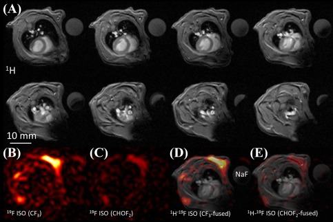
A: Multiple in vivo axial 1H MR images from one of the studied mice (eight out of ten acquired slices are shown, slice thickness = 1 mm). The 1H images correspond to the same volume as the 19F MRI. A cylindrical sodium fluoride (NaF) phantom is also visible within the field‐of‐view. B,C: Corresponding 19F images of the two ISO peaks (slice thickness = 10 mm. D,E: Merged 1H‐19F MRI of the reconstructed 19F images. For merging, a typical 1H scan was chosen within the acquired stack. 19F localizes in the skeletal muscle and fat areas of the thorax.