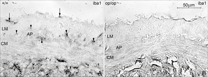
Figure 4.

iba1‐staining in the muscularis externa in ileal frozen sections from control and op/op mice. A: iba1 positive cells in control mice were present in the serosa (arrow), at the level of AP, between the longitudinal (LM), and circular muscle layer (CM; arrow head). B: In op/op mice, iba1‐immunoreactivity was absent in muscularis externa. Bar 50 μm.
