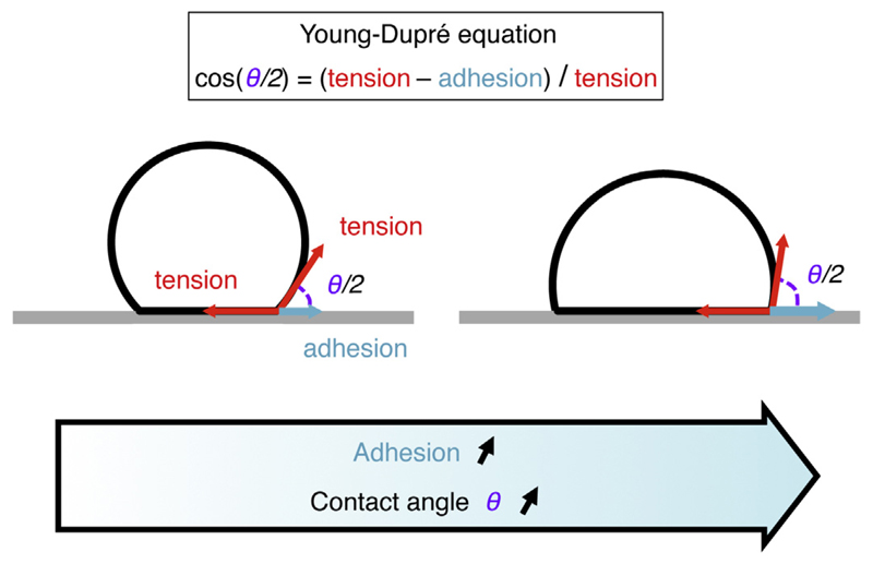
Fig. 1.
Adhesion of a droplet, vesicle or bubble to a surface. Schematic of an inert droplet, vesicle or a bubble adhering onto a surface. The adhesion can be counted in terms of tension, but it acts in the direction opposite as the surface tension along the contact. The spreading is described by the contact angle θ/2 and is governed by the Young–Dupré tension balance at the contact: cos(θ/2) = (tension – adhesion)/tension.

