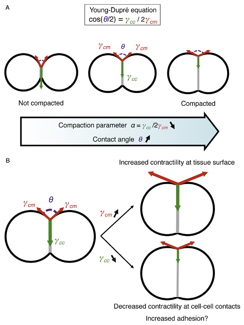
Fig. 2.
Mechanical control of compaction. A – Schematic of a minimal tissue from low (left) to high (right) compaction. The degree of compaction is given by the angles of contact θ (magenta), which results from the ratio of tension within the tissue γcc (green) and between the outside of the tissue and its surrounding γcm (red). Therefore, the compaction parameter α = γcc/2γcm = cos(θ/2), as given by the Young–Dupré equation, reflects both the degree of compaction and the balance of tensions in the tissue. Compaction corresponds to a decrease of the compaction parameter α. B – Compaction occurs when the tension γcm increases (top right) and/or the tension γcc decreases (bottom right). Contractility controls both γcm and γcc. Adhesion may theoretically control γcc directly, but its signaling to decrease contractility at cell–cell contacts constitutes the primary influence of adhesion to tissue compaction.

