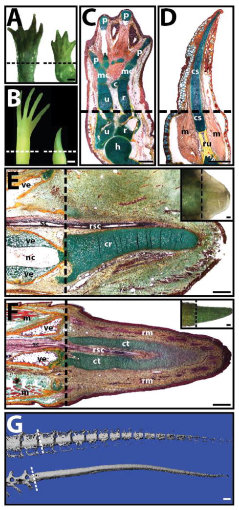
Fig. 1.
Examples of limb and tail regeneration in amphibians and lizards. (A, B) Morphological comparison of (A) salamander (Ambystoma mexicanum) and (B) frog (Xenopus laevis) forelimbs before (left) and 8 weeks after (right) amputation. Salamanders regenerate new limbs, while frogs regenerate cartilage spikes. (C, D) Histological analysis (pentachrome) of regenerated (C) salamander and (D) frog limbs. Salamanders regenerate all the skeletal elements of the upper arm and hand, while frogs regenerate a single cartilage spike. (E, F) Histological (pentachrome) and (E, F Insets) morphological analysis of (E) salamander tail 5-weeks post amputation and (F) lizard (Anolis carolinensis) tail 2 weeks post-amputation. (G) Salamander (top) and lizard (bottom) tails 10 weeks after amputation analyzed by micro computed tomography. The regenerated salamander, but not lizard, skeleton segments. Pentachrome stains cartilage green, bone orange, muscle red, and spinal cord and epidermis purple. Dashed lines denote amputation planes. c, carpal; cr, cartilage rod; cs, cartilage spike; ct, cartilage tube; h, humerus; m, muscle; mc, metacarpal; nc, notochord; p, phalanges; r, radius; rm, regenerated muscle; rsc, regenerated spinal cord; ru, radio-ulna; sc, spinal cord; u, ulna; ve, vertebra. Bar = 1 mm.

