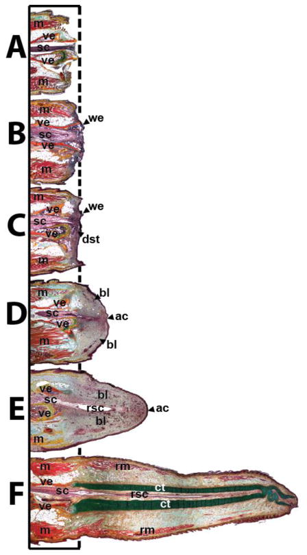
Fig. 3.
Representative lizard tails (A) 0, (B) 3, (C) 6, (D) 9, (E) 12, (F) 15, (G) and 28 days following tail loss highlighting the important structures involved with tail regrowth. ac, apical cap; bl, blastema; ct, cartilage tube; dst, degenerated stump tissue; m, muscle; rm, regenerated muscle; rsc, regenerated spinal cord; sc, spinal cord; we, wound epithelium; ve, vertebra.

