
Fig. 6.
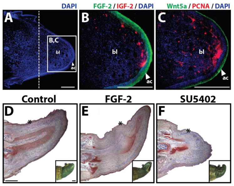
(A–C) Analysis of signaling molecules and cell proliferation markers within the regenerating lizard tail. Lizard tail blastemas (9 days following tail loss) were immunostained for IGF-2, FGF-2, Wnt5a, and PCNA. Panels B and C depict higher magnification views of region identified in Panel A. Dashed lines denote amputation planes. (D–F) Effects of FGF-2 and the FGF inhibitor SU5402 on lizard tail regeneration. Beads soaked in (D) vehicle control, (E) 100 μg/ml FGF-2, (F) or 2 mg/ml SU5402 were implanted in lizard tail blastemas. Following 7 days of growth, the tails were collected and analyzed by collagen type II immunohistochemistry. Insets of each panel depict gross tail morphology. Stars denote bead implantation sites. (D) Control tails exhibit normal ependymal tube extension. (E) Tails treated with FGF-2-soaked beads exhibit ependymal tube branches that invade toward implantation sites. (F) Tails treated with SU5402-soaked beads exhibit stunted ependymal tube invasion. ac, apical cap; bl, blastema; et, ependymal tube; ct, cartilage tube. Bar = 2 mm.