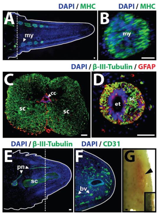
Fig. 9.
Lizard tail muscle, spinal cord, peripheral nerve, and blood vessel regeneration. (A) Lizard tail 21 days post amputation (DPA) immunostained for muscle marker myosin heavy chain (MHC). Regenerated muscles begin as distinct myomeres. (B) Close-up of a regenerated muscle myomere. (C) Original and (D) regenerated tail spinal cords analyzed immunostained for neural marker β-III-tubulin and glial cell marker glial fibrillary acidic protein (GFAP). (E, F) Lizard tail 9 DPA immunostained for (E) β-III-tubulin and (F) the blood vessel marker CD31. (E) Peripheral nerves regenerate from dorsal root ganglia in tail stumps into tail blastemas. (F) Blood vessels form at the distal lizard tail blastema tip (G) Blood vessels also form at the distal salamander tail blastema tip. Black arrowhead marks prominent vascular bed. Inset depicts gross morphology of entire salamander tail blastema. Dashed lines denote amputation planes, and solid lines trace tail outlines. bl, blastema; drg, dorsal root ganglion; pn, peripheral nerve; sc, spinal cord. Bar = 25 μm.

