Figure 1.

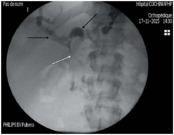
Opacification of left and right biliary ducts under fluoroscopy (black arrows) that are dilated secondary to Klatskin Bismuth III cholangiocarcinoma. No contrast opacification is seen in the main bile duct (white arrow).

Opacification of left and right biliary ducts under fluoroscopy (black arrows) that are dilated secondary to Klatskin Bismuth III cholangiocarcinoma. No contrast opacification is seen in the main bile duct (white arrow).