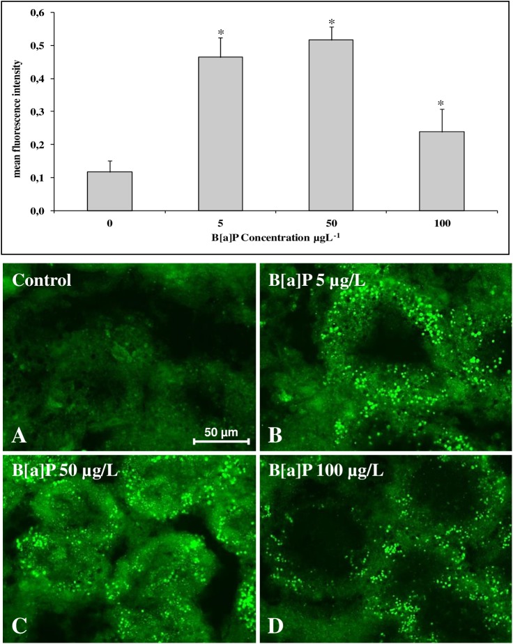
Fig 8.
(A-D) Anti-tubulin immunohistochemical staining (Chromeo™ 488 conjugated secondary antibody) of digestive gland tissue sections from mussels exposed to different experimental conditions (A = Control; B = B[a]P 5 μg/L; C = B[a]P 50 μg/L; D = B[a]P 100 μg/L). (E) Quantitative fluorescence analysis of anti-tubulin immunoreaction. Data are mean ± SD of at least five replicates; * = p < 0.05 (Mann-Whitney U-test).

