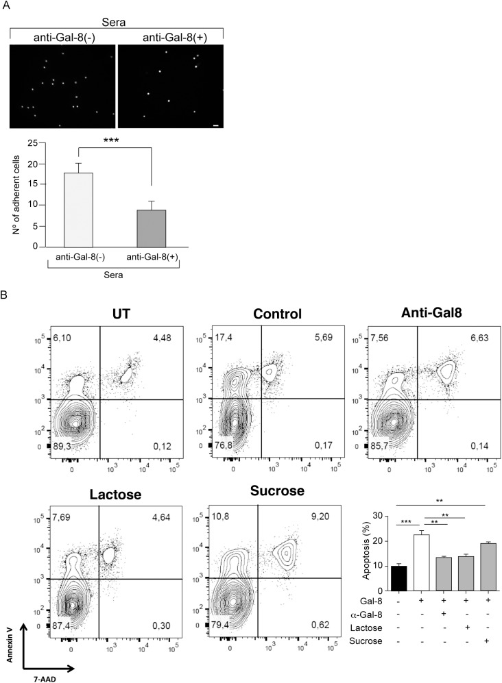
Fig 8. Function-blocking activity of anti-Gal-8 autoantibodies.
(A) Anti-Gal-8(+) sera block the adhesion of PBMC to Gal-8-coated coverslips. Graph shows number of adhered cells (Average ± SE of three anti-Gal-8(-) and three anti-Gal-8(+) sera tested in triplicate) (***p<0.001; Student’s t-test). (B) Anti-Gal-8 autoantibodies inhibit Gal-8-induced apoptosis of Th17 cells. In vitro differentiated Th17 cells from IL-17A-GFP reporter mice were purified based on IL-17A expression (GFP+) and incubated with Gal-8 (20 μg/ml) in the presence of lactose, sucrose or anti-Gal-8 antibodies affinity purified from pooled serum of MS patients. The extent of apoptosis was quantified as the frequency of Annexin V+ 7AAD+ cells of the sample relative to the frequency of Annexin V+ 7AAD+ cells of the untreated control. Representative contour plots are shown in upper panels. Quantification of a representative experiment is shown in the lower panel. Values represent mean + SEM of triplicates. Data from a representative from four independent experiments is shown. **, p<0.01; ***, p < .001 by one-way ANOVA followed by Tukey’s post-hoc test.

