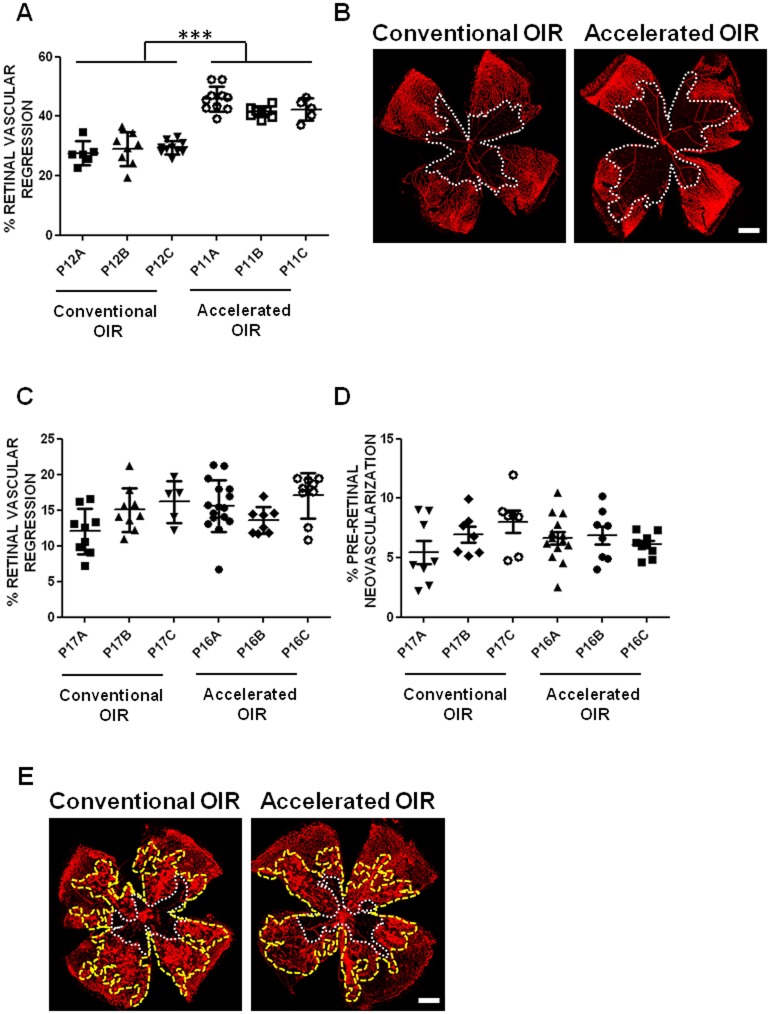
Fig 1. Retinal vasculature regression and pre-retinal neovascularization following hyperoxia and 5 days in room air.
(A) The extent of oxygen-induced retinal vascular regression was consistently greater (in 3 independent experimental groups) in the accelerated protocol (85% O2 from P8 to P11) than in the conventional protocol (75% O2 from P7 to P12). No significant differences were evident between independent experimental groups within each protocol, and the variances were similar (Barlett's test, p = 0.6429 for retinal vascular regression and p = 0.1415 for pre-retinal neovascularization). (B) Representative images show the extent of retinal vascular regression (delineated in white) in isolectin B4-stained flat-mounted retinas of mice in the conventional and the accelerated OIR protocols. Five days following return to room air, the extents of both persistent retinal vascular regression (C) and pre-retinal neovascularization (D) were similar in retinas of mice in both protocols. (E) Representative images of flat-mounted retinas of mice from the conventional and accelerated protocols illustrate the area of persistent retinal vascular regression (delineated in white), and the area of pre-retinal neovascularization (delineated in yellow). Scale bars: 0.5 mm. n = 6–14 per group. Data are expressed as means ± SEM.

