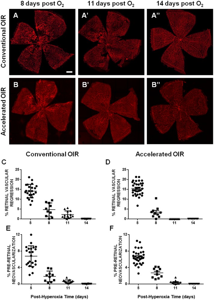
Fig 2. Time courses of retinal vasculature regeneration and pre-retinal neovascular regression.
(A, B) Representative flat-mounted retinas from mice exposed to conventional OIR (75% O2, upper panel) or accelerated OIR (85% O2, lower panel) at 8 (A, B), 11 (A’, B’) and 14 days after the end of hyperoxia (A”,B”), showing progressive retinal vascular regeneration, and regression of the neovascular tufts. (C, D) Analysis of the persistent retinal vascular regression demonstrated more rapid vascular regeneration in the accelerated protocol at 11 days post-hyperoxia (p = 0.015). (E, F) No differences were found in the kinetics of regression of neovascular lesions between the two experimental conditions. Scale bars: 0.4 mm. n = 7–13 per group. Data are expressed as means ± SEM.

